A Breakthrough in Oocyte Selection for IVF
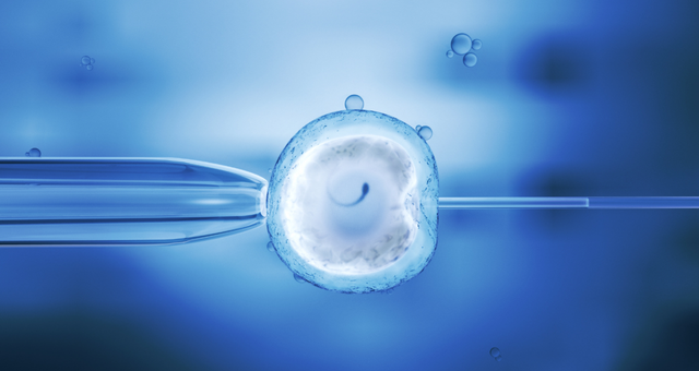
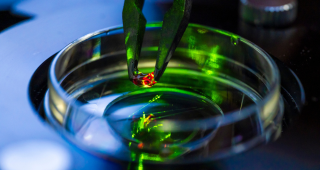
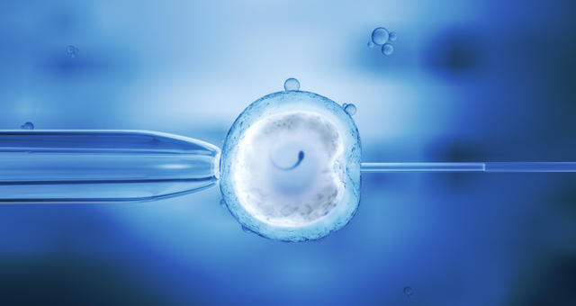 Polish scientists have become the first in the world to use holotomography for the three-dimensional imaging and assessment of live oocytes. The method makes it possible to determine the developmental potential of oocytes before in vitro fertilisation.
This is a breakthrough that could, on one hand, increase the effectiveness of infertility treatment by improving the chances of embryo implantation, carrying the pregnancy to term, and giving birth to a healthy child. On the other hand, it could reduce the number of a woman′s oocytes being fertilized, thereby decreasing the number of so-called surplus embryos-specifically those that are not transferred to the uterus during the current treatment cycle.
The innovative solution has been developed by a team of scientists from the Małopolska Centre of Biotechnology at Jagiellonian University, working in collaboration with the Institute of Nuclear Physics of the Polish Academy of Sciences. The technology enables rapid three-dimensional visualisation of the lipid profile of live oocytes, providing a reliable assessment of their metabolic status. This makes it possible to identify oocytes with the greatest developmental potential and, ultimately, improve success rates in IVF procedures.
The holotomography-based method represents a major qualitative leap in this field. Instead of relying on a subjective assessment of morphology, embryologists gain access to objective data reflecting the actual physiological and biochemical condition of the cell. This enables them to identify oocytes offering the highest likelihood of successful implantation of the embryo and the healthy pregnancy.
“Two oocytes that appear identical under a microscope may have completely different developmental potential. This is influenced, among other factors, by the volume of lipids accumulated within the cells, as well as their distribution and structure. Our method allows us to look inside live oocytes and assess their condition, enabling appropriate selection. For the first time, an embryologist can select oocytes with optimal metabolic parameters for fertilisation, thereby increasing the chances of embryo implantation, a healthy pregnancy and the birth of a healthy child”, says Professor Grażyna Ptak of the Małopolska Centre of Biotechnology at Jagiellonian University, who originated the concept behind the method.
The most important benefit of the new solution is the increased likelihood of achieving a pregnancy resulting in the birth of a healthy child. Potentially, the method could also reduce the risk of cardiometabolic diseases later in life – an issue observed in some children born as a result of assisted reproductive technologies and supported by studies using animal models. The technology could therefore contribute to improving the long-term success of IVF procedures.
The technique allows researchers to visualise the interior of an individual living cell without using dyes or interfering with the biological material. It is therefore entirely non-invasive.
In the technology developed by the research team, scientists analyse the oocyte’s lipid profile – that is, the distribution and volume of lipid structures throughout the entire cell. Assessing the condition of lipids makes it possible to detect adverse oxidative processes before damage to proteins or the genetic material within the cell occurs. Lipids are among the first cellular components to respond to oxidative stress, which induces changes in membrane structure and consequently reduces the developmental competence of the oocyte.
“Holotomography allows us to visualise a living cell in three dimensions without using any labels or markers. We analyse the distribution of lipids throughout the entire volume of the oocyte, obtaining information that conventional light microscopy simply cannot provide”, explains Joanna Depciuch-Czarny of the Institute of Nuclear Physics of the Polish Academy of Sciences, a co-developer of the new technology.
The examination involves generating a computer-based three-dimensional reconstruction that enables quantitative analysis of cellular parameters and the selection and ranking of oocytes – from those with the most favourable characteristics to those displaying the greatest abnormalities. The entire procedure takes approximately ten minutes.
As scientists and clinicians emphasise, all existing systems for evaluating oocytes solely on the basis of visual assessment – including those using artificial intelligence – are unable to determine oocyte developmental competence effectively. There is broad agreement that, apart from clearly visible morphological abnormalities, selecting oocytes on the basis of morphology does not improve pregnancy rates compared with procedures in which such morphological selection is not performed.
Until now, holotomography has primarily been used to analyse cells adhering to a substrate. The research team developed a solution enabling three-dimensional analysis of oocytes, which remain suspended in the culture medium. Imaging is performed at the nanometre scale, providing a very high level of precision. This aspect of the technology constitutes one of the key elements covered by the patent application.
“Developing our method required us to solve a number of technical challenges associated with sample preparation. One of them was finding an effective way to image a single cell suspended in culture medium using a holotomographic microscope. Another challenge was immobilising an individual cell that does not adhere to a substrate. We are pioneers in this area”, says Hafsa Gulzar, a co-developer of the technology.
Another challenge successfully addressed by the researchers was the optimisation of all preparation and analytical steps so that a pool of retrieved oocytes could be examined and analysed in 3D within ten minutes. This timeframe makes the method suitable for implementation in a clinical setting.
 “The technology addresses a clear need for effective methods of oocyte assessment. We are currently analysing potential commercialisation pathways and supporting the research team in identifying partners interested in the further development of the solution, with the ultimate goal of enabling its use in embryology laboratories”, says Dr Gabriela Konopka-Cupiał, Director of the Centre for Technology Transfer CITTRU at Jagiellonian University.
The research team is currently pursuing research and validation activities along three parallel paths. The first involves comprehensive studies using animal models, providing information on the long-term effects of changes in oocyte lipid profiles on the health of adult offspring.
The second focuses on characterising the causes of alterations in the lipid profiles of oocytes made available for research by collaborating fertility clinics.
In parallel, work is under way on an artificial intelligence component based on machine learning. Its purpose is to standardise and accelerate the analysis of data obtained through holotomography. Ultimately, this component is expected to become an integral part of the software used with holotomographic microscopes, enabling embryologists to perform rapid and objective assessments of oocyte quality.
“Despite the rapid development of assisted reproductive technologies, the effectiveness of assisted reproduction technologies– still remains at only around 35%. One of the main factors limiting success rates is the lack of an objective and reliable method for assessing the quality of oocytes selected for fertilisation. I believe that our technology will drive progress in this field and help solve a problem that fertility clinics have been facing for many years”, adds Professor Grażyna Ptak.
The project was carried out by a team led by Professor Grażyna Ptak of the Małopolska Centre of Biotechnology at Jagiellonian University (MCB JU); Hafsa Gulzar, a doctoral researcher at MCB JU working under Professor Ptak’s supervision; and Joanna Depciuch-Czarny of the Institute of Nuclear Physics of the Polish Academy of Sciences.
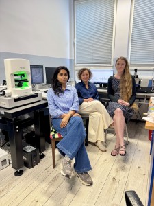
On the photo, from the left: mgr Hafsa Gulzar i prof. dr hab. Grażyna Ptak (Małopolskie Centrum Biotechnologii UJ) oraz dr hab. inż. Joanna Depciuch-Czarny (Instytut Fizyki Jądrowej PAN).
The innovative solution has been developed by a team of scientists from the Małopolska Centre of Biotechnology at Jagiellonian University, working in collaboration with the Institute of Nuclear Physics of the Polish Academy of Sciences. The technology enables rapid three-dimensional visualisation of the lipid profile of live oocytes, providing a reliable assessment of their metabolic status. This makes it possible to identify oocytes with the greatest developmental potential and, ultimately, improve success rates in IVF procedures.
A New Standard in Oocyte Assessment
Despite the tremendous progress made in reproductive medicine, no effective method for assessing oocyte developmental competence has yet been introduced into IVF practice. To this day, the selection of an oocyte for fertilisation is based primarily on its appearance under a microscope. The problem is that most oocytes look almost identical, even though they may differ dramatically in terms of their metabolism and internal structure – and, consequently, in their ability to develop successfully after fertilisation.The holotomography-based method represents a major qualitative leap in this field. Instead of relying on a subjective assessment of morphology, embryologists gain access to objective data reflecting the actual physiological and biochemical condition of the cell. This enables them to identify oocytes offering the highest likelihood of successful implantation of the embryo and the healthy pregnancy.
“Two oocytes that appear identical under a microscope may have completely different developmental potential. This is influenced, among other factors, by the volume of lipids accumulated within the cells, as well as their distribution and structure. Our method allows us to look inside live oocytes and assess their condition, enabling appropriate selection. For the first time, an embryologist can select oocytes with optimal metabolic parameters for fertilisation, thereby increasing the chances of embryo implantation, a healthy pregnancy and the birth of a healthy child”, says Professor Grażyna Ptak of the Małopolska Centre of Biotechnology at Jagiellonian University, who originated the concept behind the method.
The most important benefit of the new solution is the increased likelihood of achieving a pregnancy resulting in the birth of a healthy child. Potentially, the method could also reduce the risk of cardiometabolic diseases later in life – an issue observed in some children born as a result of assisted reproductive technologies and supported by studies using animal models. The technology could therefore contribute to improving the long-term success of IVF procedures.
Lipids Hold the Key to Oocyte Health
The new method uses holotomography – an advanced 3D imaging technique based on measuring the refractive index of structures within the material being examined.The technique allows researchers to visualise the interior of an individual living cell without using dyes or interfering with the biological material. It is therefore entirely non-invasive.
In the technology developed by the research team, scientists analyse the oocyte’s lipid profile – that is, the distribution and volume of lipid structures throughout the entire cell. Assessing the condition of lipids makes it possible to detect adverse oxidative processes before damage to proteins or the genetic material within the cell occurs. Lipids are among the first cellular components to respond to oxidative stress, which induces changes in membrane structure and consequently reduces the developmental competence of the oocyte.
“Holotomography allows us to visualise a living cell in three dimensions without using any labels or markers. We analyse the distribution of lipids throughout the entire volume of the oocyte, obtaining information that conventional light microscopy simply cannot provide”, explains Joanna Depciuch-Czarny of the Institute of Nuclear Physics of the Polish Academy of Sciences, a co-developer of the new technology.
The examination involves generating a computer-based three-dimensional reconstruction that enables quantitative analysis of cellular parameters and the selection and ranking of oocytes – from those with the most favourable characteristics to those displaying the greatest abnormalities. The entire procedure takes approximately ten minutes.
Fertility Clinics Have Been Waiting for a Solution Like This
The new technology has generated considerable interest among fertility clinics. Centres performing IVF procedures are looking for solutions that can improve treatment outcomes and reduce the number of unsuccessful attempts. Ultimately, the technology could become part of embryologists’ routine workflow, supporting their decisions with objective data analysis and replacing the limitations of visual assessment.As scientists and clinicians emphasise, all existing systems for evaluating oocytes solely on the basis of visual assessment – including those using artificial intelligence – are unable to determine oocyte developmental competence effectively. There is broad agreement that, apart from clearly visible morphological abnormalities, selecting oocytes on the basis of morphology does not improve pregnancy rates compared with procedures in which such morphological selection is not performed.
A Breakthrough in Holotomography Itself
The research team’s achievement goes beyond simply applying holotomography to oocyte assessment. An equally important advance was the development of a proprietary method for preparing oocytes for examination and enabling their complete three-dimensional imaging.Until now, holotomography has primarily been used to analyse cells adhering to a substrate. The research team developed a solution enabling three-dimensional analysis of oocytes, which remain suspended in the culture medium. Imaging is performed at the nanometre scale, providing a very high level of precision. This aspect of the technology constitutes one of the key elements covered by the patent application.
“Developing our method required us to solve a number of technical challenges associated with sample preparation. One of them was finding an effective way to image a single cell suspended in culture medium using a holotomographic microscope. Another challenge was immobilising an individual cell that does not adhere to a substrate. We are pioneers in this area”, says Hafsa Gulzar, a co-developer of the technology.
Another challenge successfully addressed by the researchers was the optimisation of all preparation and analytical steps so that a pool of retrieved oocytes could be examined and analysed in 3D within ten minutes. This timeframe makes the method suitable for implementation in a clinical setting.
Patent Protection and the Next Stage of Development
The researchers are confident that the technology represents a new direction for diagnostic development in reproductive medicine. A patent application covering the method has been filed in Poland, and the solution has substantial commercialisation potential. The scientists envisage integrating the technology with holotomographic microscopes for use by fertility clinics.“The technology addresses a clear need for effective methods of oocyte assessment. We are currently analysing potential commercialisation pathways and supporting the research team in identifying partners interested in the further development of the solution, with the ultimate goal of enabling its use in embryology laboratories”, says Dr Gabriela Konopka-Cupiał, Director of the Centre for Technology Transfer CITTRU at Jagiellonian University.The research team is currently pursuing research and validation activities along three parallel paths. The first involves comprehensive studies using animal models, providing information on the long-term effects of changes in oocyte lipid profiles on the health of adult offspring.
The second focuses on characterising the causes of alterations in the lipid profiles of oocytes made available for research by collaborating fertility clinics.
In parallel, work is under way on an artificial intelligence component based on machine learning. Its purpose is to standardise and accelerate the analysis of data obtained through holotomography. Ultimately, this component is expected to become an integral part of the software used with holotomographic microscopes, enabling embryologists to perform rapid and objective assessments of oocyte quality.
“Despite the rapid development of assisted reproductive technologies, the effectiveness of assisted reproduction technologies– still remains at only around 35%. One of the main factors limiting success rates is the lack of an objective and reliable method for assessing the quality of oocytes selected for fertilisation. I believe that our technology will drive progress in this field and help solve a problem that fertility clinics have been facing for many years”, adds Professor Grażyna Ptak.
The project was carried out by a team led by Professor Grażyna Ptak of the Małopolska Centre of Biotechnology at Jagiellonian University (MCB JU); Hafsa Gulzar, a doctoral researcher at MCB JU working under Professor Ptak’s supervision; and Joanna Depciuch-Czarny of the Institute of Nuclear Physics of the Polish Academy of Sciences.
On the photo, from the left: mgr Hafsa Gulzar i prof. dr hab. Grażyna Ptak (Małopolskie Centrum Biotechnologii UJ) oraz dr hab. inż. Joanna Depciuch-Czarny (Instytut Fizyki Jądrowej PAN).